Georgia Tech's School of Chemical and Biomolecular Engineering (ChBE) has announced the winners of its first-ever Stunning Science in ChBE Image Contest, which was open to all current ChBE students (undergraduate and graduate) and postdoctoral researchers in ChBE labs.
"With over 30 entries representing 12 different ChBE labs, the response to this contest highlights the diversity of research being performed in our School. The six undergrads, 15 grad students, and 11 postdocs and research scientists who entered amazed us with their creative images and compelling captions showcasing the truly innovative work that is ongoing in ChBE," said Jacqueline Snedeker, the chair of ChBE's Communications Committee who is a senior academic professional (director of the Technical Communications Program).
Submitted images could depict and object or phenomenon from ChBE obtained via such means as: Microscopy, Photographs (e.g., of 3-D printed objects), Medical imaging, Schematics, Spectra, Chromatographs, Simulation results, and so on. Photos could be featured on the ChBE website and social media and framed to hang on building walls.
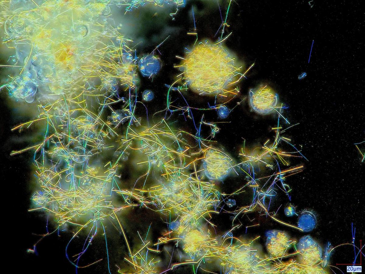
The first place winner is Grace Henderson, who will receive $500. She is an undergraduate student working in Associate Professor Michael Filler's laboratory. Her image and caption appear below.
Caption: Nanowire-filled hollow silica spheres, referred to in our lab as geodes, exhibit a captivating cosmic display when observed with a Keyence Digital Microscope. These geodes display vivid colors that are perceptible to the unaided eye, a result of the optical properties inherent to nanowires. The colors of the nanowires correspond to their diameters, which can be controlled by utilizing distinct sizes of nanowire gold catalysts. The light scattering attributes of these geodes have potential use in pigments that possess electronic functionalities.
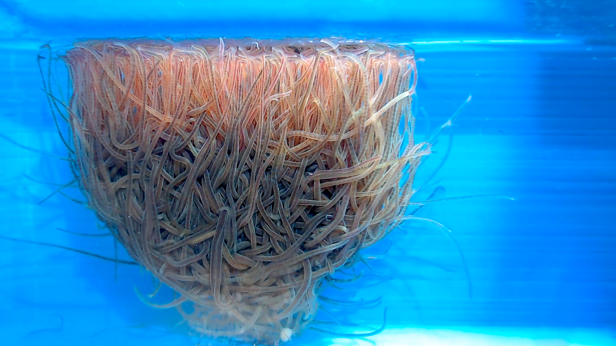
The second -place winner is Harry Tuazon, who will receive $300. He is a graduate student and member of Assistant Professor Saad Bhamla's lab. His image and caption appear below.
Caption: This image depicts a large collective of Lumbriculus variegatus (blackworms) at the air-water interface floating like a buoy. This “worm buoy” was made possible by interfacial surface tension, which allows them to latch and lift each other up, forming a floating entangled structure. The structure is formed not only for interfacial latching but also for respiration, as blackworms can supplement respiration using their tails to obtain oxygen directly from air. The buoy is a function of population dynamics and the amount of dissolved oxygen, allowing worms to breathe better together and entangle stronger with one another. Aside from personally discovering this phenomenon in the Saad Bhamla Lab, it has important implications to soft matter physics and biophysics, as it provides insights into the dynamics of collective animal behavior. Furthermore, it has potential applications in swarm robotics and the design of artificial systems that can adapt and respond to changing environments.
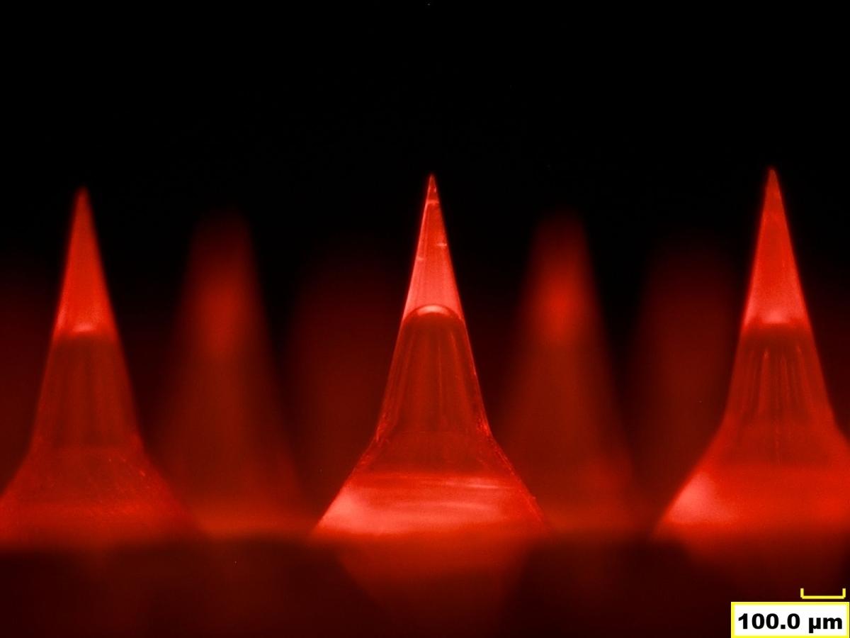
The third-place winner is Erkan Azizoglu, who will receive $150. He is a research scientist in Regents' Professor and Regents' Entrepreneur Mark Prausnitz's lab. His image and caption appear below.
Caption: Hollow but mighty: Microneedles are minimally invasive structures used for drug delivery, diagnostics, and vaccine administration. They penetrate the skin's outermost layer with minimal pain or discomfort. Rapid separation of microneedle tips is essential to deliver the cargo immediately and efficiently. Hollow microneedles are fabricated by entrapping air between the tip and the base. The image captured by a digital microscope (Hirox, KH 8700) shows the uniform distribution of the air in the microneedles. The hollow body acts like a bubble, and the insertion force causes them to pop, allowing the cargo to be detached and left in the skin. This method reduces the application time to mere seconds, making it a user-friendly option for healthcare professionals and patients.
The contest judging panel included ChBE faculty members Alex Abramson, Victor Breedveld, Martha Grover, and Jacqueline Snedeker, as well as Georgia Tech communications staff members Brad Dixon, Laurie Haigh, Candler Hobbs, and Jason Maderer. From their rankings, three Honorable Mentions were named.
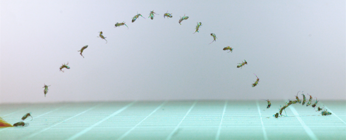
An Honorable Mention goes to Jacob Harrison, a postdoctoral researcher in Assistant Professor Saad Bhamla's lab. His image and caption appear below.
Caption: A slender springtail (Pogonognathellus negritus) leaping off the ground. The frames were taken from a high-speed video filmed at 4,000 frames per second and then stacked to show a full trajectory jump. This springtail is only a couple of millimeters long but can produce movements faster than the blink of an eye. Highspeed videography provides a window into the realm of the ultrafast behaviors that are too fast to see with our own eyes.
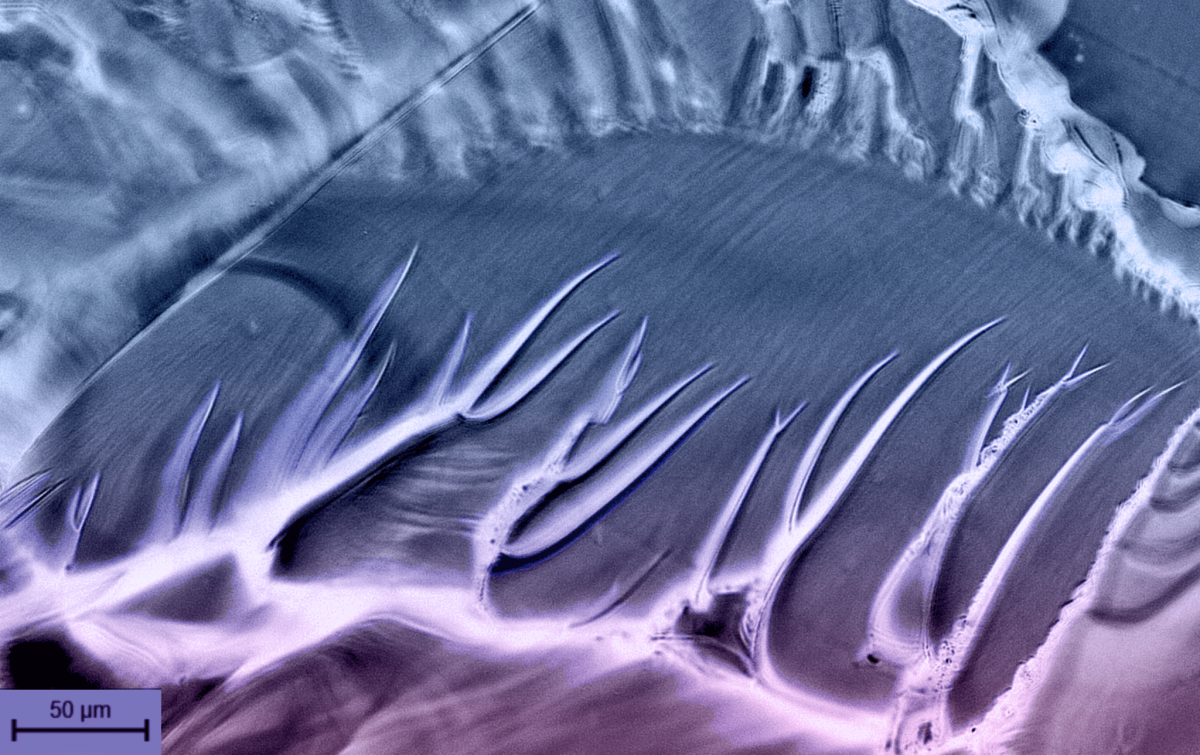
An Honorable Mention also goes to Alexandra Marnot, a graduate student and member of Assistant Professor Blair Brettmann's lab. Her image and caption appear below.
Caption: Fracture propagation through a UV crosslinked polymer mixture. Microscope image, colorized. Fracture paths occur because the crosslinking density between monomers within the mixture is very high, therefore the material cannot swell easily when placed in a solvent. Crosslinking density of polymer mixtures is important in dictating the cured material's mechanical properties, such as elongation and toughness.
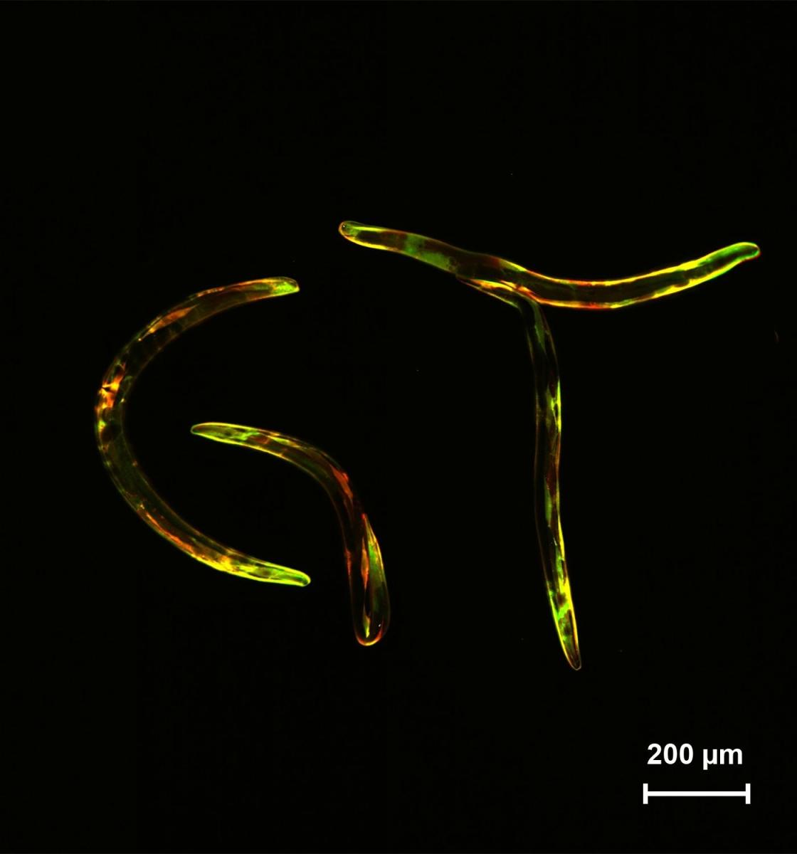
An Honorable Mention also goes to Hyun Jee Lee, a graduate student and member of Professor Hang Lu's lab. Her photo and caption appear below.
Caption: The image depicts four microscopic nematode worms (C. elegans) that were genetically modified to express red and green fluorescent proteins in their muscles. Fun fact, these worms are only about the size of a printed comma “,”. To make this image, the nematodes were arranged to spell “GT” and imaged using two color fluorescence confocal microscopy. The combination of red and green allows optical measurement of muscle activation; the green becomes brighter when the muscle activates, and the red enables automated detection of the muscles by computer vision algorithms. Muscle activation in genetically modified C. elegans enables investigation of genetic mechanisms that coordinate neuron and muscle activities in response to changing environmental conditions, how these mechanisms might break down during neurodegenerative diseases, and how we might adapt them towards bioinspired design of robots that more efficiently navigate through complex terrain using flexible mechanics and feed-forward control.
Contact: Brad Dixon